A kneecap dislocation occurs when the patella slips out of its normal position, usually towards the outer side of the knee.
Common causes include sudden twisting movements, sports injuries, falls and weak supporting muscles.
Symptoms include severe knee pain, swelling, bruising and difficulty walking or bending the knee.
Treatment may involve rest, a knee brace, pain relief and physiotherapy. Surgery may be needed for repeated dislocations or significant damage.
Strengthening the thigh and hip muscles, improving balance and following physiotherapy can help prevent recurrent kneecap dislocations.
A kneecap dislocation occurs when the patella (kneecap) slips out of its normal position, usually towards the outer side of the knee. It can happen due to a sudden twist, a sports injury, a fall or an awkward landing. The injury can cause severe knee pain, swelling and difficulty walking. While the kneecap may sometimes return to its normal position on its own, proper medical evaluation and rehabilitation are important to prevent another dislocation.
If your kneecap keeps slipping or you have experienced a displaced kneecap before, understanding why it happens and how to protect your knee can help you avoid repeated injuries.
The patella is a small, triangular bone located at the front of your knee. It protects the knee joint and helps the thigh muscles straighten your leg. It normally moves up and down within a groove at the lower end of the thigh bone (femur) when you bend or straighten your knee.
A patella dislocation occurs when the kneecap moves completely out of this groove. In most cases, it slips towards the outer side of the knee. This is also known as a lateral patellar dislocation.
A partial movement, where the kneecap moves out of its normal position but does not completely leave the groove, is called patellar subluxation.
A displaced patella can sometimes return to its normal position without medical assistance. However, this does not mean the injury has healed completely. The ligaments and cartilage around the kneecap may still be damaged.
It is also important to understand that a dislocated kneecap is different from a dislocated knee joint. A knee joint dislocation involves the thigh bone and shin bone losing their normal alignment. It is a more serious injury that can damage blood vessels and nerves and requires emergency medical attention.
A kneecap dislocation usually happens when the knee twists or turns suddenly while the foot remains firmly on the ground. However, some people are more likely to experience this injury because of their knee structure or ligament strength.
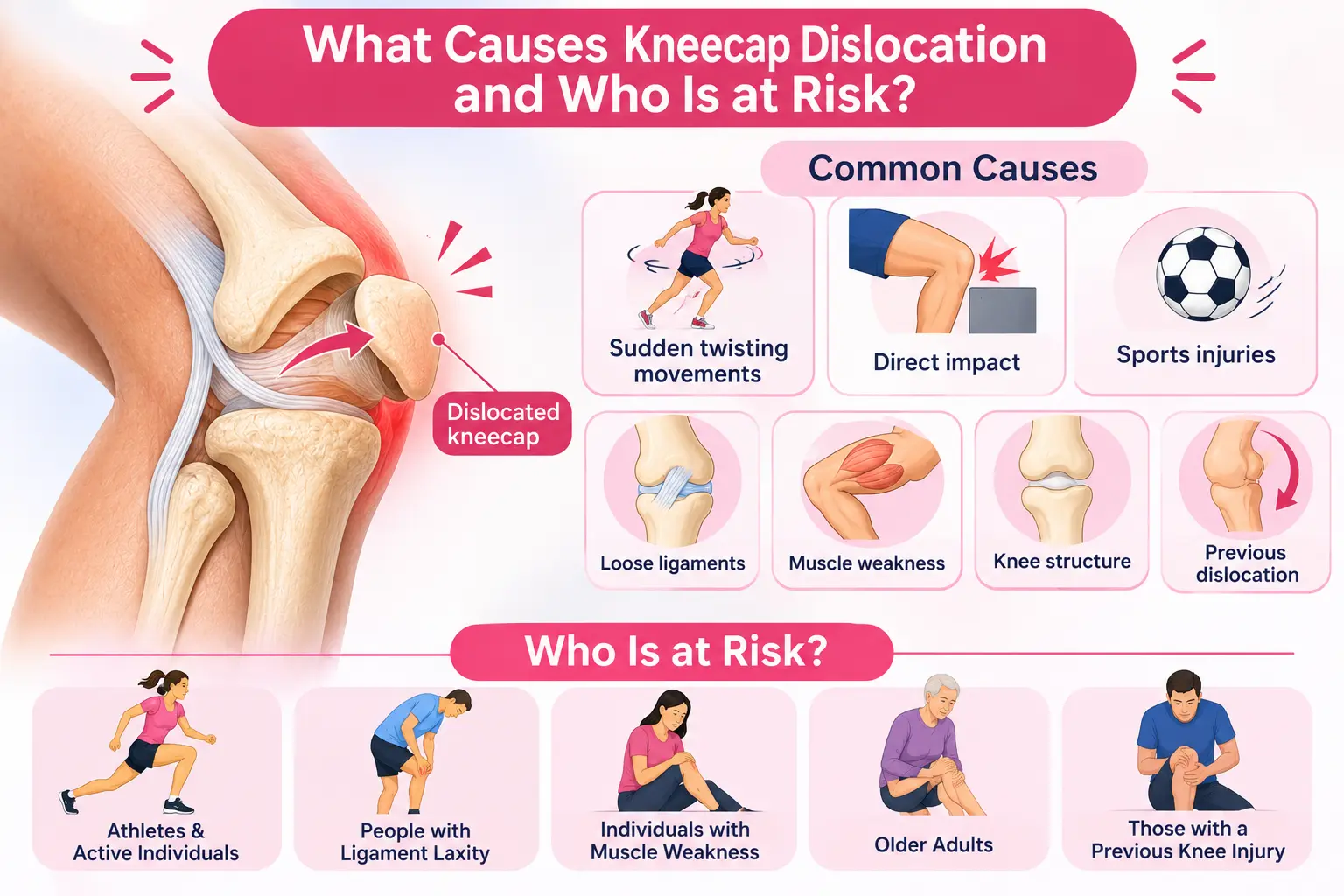
Common causes and risk factors include:
Some people also have congenital patellar dislocation, a condition in which the kneecap is displaced from birth due to differences in the development or structure of the knee. This is different from an injury-related dislocation and may require specialised treatment.
The symptoms of a dislocated kneecap usually appear immediately after the injury. Their severity depends on how far the patella has moved and whether the surrounding ligaments or cartilage are damaged.
Common dislocated knee symptoms include:
Sometimes, a slipped patella returns to its normal position before you reach a doctor. Even then, pain, swelling and a feeling of instability may continue.
Do not try to force a displaced kneecap back into place yourself. Seek medical attention, particularly if you cannot move your knee, have severe pain or notice significant swelling.
Doctors diagnose a kneecap dislocation through a physical examination and imaging tests. Treatment depends on whether it is the first dislocation, whether the kneecap has returned to its normal position and whether there is damage to the surrounding structures.
Your doctor will examine the knee to check its position, swelling, tenderness and movement. They may also assess the stability of the kneecap and compare the affected knee with the other one.
Imaging tests may include:
These tests help doctors decide whether non-surgical treatment is sufficient or whether surgery may be needed.
Most first-time kneecap dislocations without major associated injuries can be managed without surgery.
Treatment options may include:
Surgery may be recommended if the kneecap dislocates repeatedly, there is significant cartilage or bone damage, or the knee remains unstable despite appropriate rehabilitation.
Depending on the injury, surgical options may include medial patellofemoral ligament (MPFL) reconstruction to restore the ligament that helps prevent the kneecap from slipping towards the outer side. In some patients, doctors may also recommend procedures to correct underlying bone alignment or structural problems.
The choice of surgery depends on the patient's age, knee anatomy, injury severity and history of previous dislocations.
If your kneecap has dislocated once, you may have a higher risk of experiencing it again. This is because the ligaments that support the patella may have been stretched or damaged during the first injury.
Recurrent subluxation of patella can also occur when the kneecap repeatedly moves partially out of its normal position. The good news is that appropriate rehabilitation and changes in physical activity can help improve knee stability.
Here are some ways to reduce the risk of another dislocation:
If your knee cap keeps slipping despite physiotherapy, consult an orthopaedic specialist. Repeated dislocations may indicate an underlying structural problem that needs further evaluation.
A kneecap dislocation can cause sudden pain, swelling and difficulty moving your knee. Although many first-time injuries can be treated without surgery, ignoring the injury or returning to physical activities too soon can increase the risk of another episode.
Early diagnosis, appropriate treatment and a guided physiotherapy programme can help restore knee stability. If your kneecap keeps slipping or you experience repeated dislocations, consult an orthopaedic specialist to identify the cause and discuss suitable treatment options.
The kneecap may keep dislocating because the supporting ligaments have become stretched or damaged after an earlier injury. Other factors include weak thigh muscles, a shallow groove in the thigh bone, a high-riding kneecap or poor knee alignment. Repeated episodes need an orthopaedic assessment to identify the underlying cause.
Yes, many first-time kneecap dislocations can heal without surgery, especially when there is no major bone or cartilage injury. Treatment usually involves reducing the kneecap if needed, using a brace for support and following a physiotherapy programme. Surgery may be considered for repeated dislocations or significant associated injuries.
Some people can walk after a kneecap dislocation, particularly if the patella has returned to its normal position. However, walking may be painful or difficult due to swelling and ligament damage. Avoid putting unnecessary weight on the injured leg and seek medical evaluation.
Yes, a kneecap dislocation can increase the risk of developing knee joint problems, including patellofemoral osteoarthritis. This is more likely when the injury damages the cartilage behind the kneecap or when repeated dislocations cause ongoing joint damage.
Exercises such as quadriceps strengthening, straight-leg raises, gentle knee extensions and hip strengthening may help improve knee stability. Balance exercises may also be included during later recovery. However, exercises should begin only when recommended by your doctor or physiotherapist, as the right programme depends on the severity of the injury.
Physiotherapy can help reduce the risk of another dislocation by strengthening the thigh and hip muscles, improving balance and correcting movement patterns. However, it may not fully prevent recurrence in people with significant structural abnormalities or severe ligament damage.
Yes. When the kneecap slips out of its normal position, it can stretch or tear the supporting ligaments, particularly the medial patellofemoral ligament (MPFL). It may also damage the cartilage behind the kneecap or cause small bone and cartilage fragments to break away. An MRI may be recommended to assess these injuries.
Written and Verified by:

Dr. Rakesh Rajput is the HOD & Director of Orthopaedics Dept. at CMRI, Kolkata, with over 25 years of experience. He specializes in robotic knee and hip replacement, joint preservation, complex trauma care, and revision surgeries.
Similar Orthopedics & Joint Replacement Blogs
Book Your Appointment TODAY
© 2024 CMRI Kolkata. All Rights Reserved.