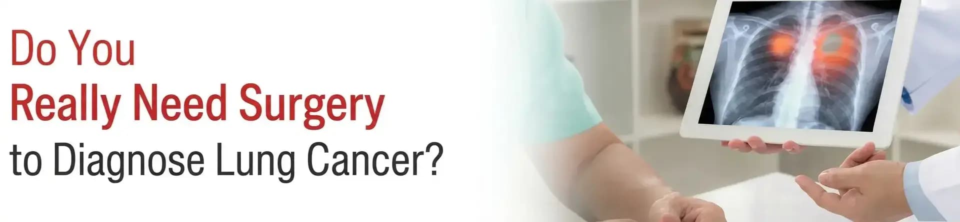
A suspicious lung nodule does not always require open surgery for diagnosis. PET-CT, advanced bronchoscopy and EBUS can help doctors diagnose and stage lung cancer while obtaining tissue samples through minimally invasive techniques.
“When a CT scan detects a suspicious lung nodule, patients often fear that open surgery will be required simply to establish the diagnosis. Fortunately, patients can now be evaluated through minimally invasive techniques. Not every nodule is cancer or requires immediate biopsy; its size, appearance, growth, location and the patient’s risk determine whether surveillance or tissue sampling is appropriate.
PET-CT adds another important layer. It shows the metabolic activity of the nodule, lymph nodes and possible disease elsewhere in the body, helping us choose the most informative site for biopsy. However, PET-CT cannot by itself confirm cancer because infection and inflammation may also show increased uptake. Tissue diagnosis remains essential.
The key advantage of the bronchoscopic pathway is that it can provide diagnosis and staging during the same procedure. We can sample a peripheral lung nodule using a thin or ultrathin bronchoscope with radial-probe EBUS guidance and, when indicated, use convex-probe EBUS in the same sitting to sample mediastinal or hilar lymph nodes. This can reduce the need for separate procedures and provide the information required for treatment planning.
At CK Birla Hospitals - RBH Jaipur, we have standard, thin and ultrathin bronchoscopes, radial-probe and convex-probe EBUS, along with a 1.1-mm cryoprobe. For peripheral nodules, we study the CT images beforehand and perform cognitive bronchial mapping or bronchial branch tracing to plan the route through progressively smaller airways. Radial EBUS helps confirm proximity to the target, while forceps, needle or cryobiopsy can be used to obtain tissue. Cryobiopsy may provide a larger, well-preserved sample for histological and molecular testing.
The 2025 VERITAS randomized trial, which used electromagnetic navigation with integrated digital tomosynthesis, strengthened the role of advanced navigational bronchoscopy. For 10 - 30 mm peripheral nodules, diagnostic accuracy was noninferior to CT-guided transthoracic biopsy - 79.0% versus 73.6% - while pneumothorax occurred much less often, 3.3% versus 28.3%. It supports the safety advantage of approaching suitable nodules through the airways.
Our objective is a coordinated pathway - from PET-CT assessment to minimally invasive diagnosis, nodal staging and adequate tissue acquisition - while avoiding open surgery whenever safely possible.”
Written and Verified by:
.webp&w=256&q=75)
Dr. Rakesh Godara is Additional Director of Pulmonology Dept. at CK Birla Hospital, Jaipur with over 18 years of experience. He specializes in ARDS, bronchoscopic management of hemoptysis, central airway obstruction, endobronchial ultrasound, and medical thoracoscopy/pleuroscopy.
Similar Pulmonology Blogs
Book Your Appointment TODAY
© 2024 RBH Jaipur. All Rights Reserved.


