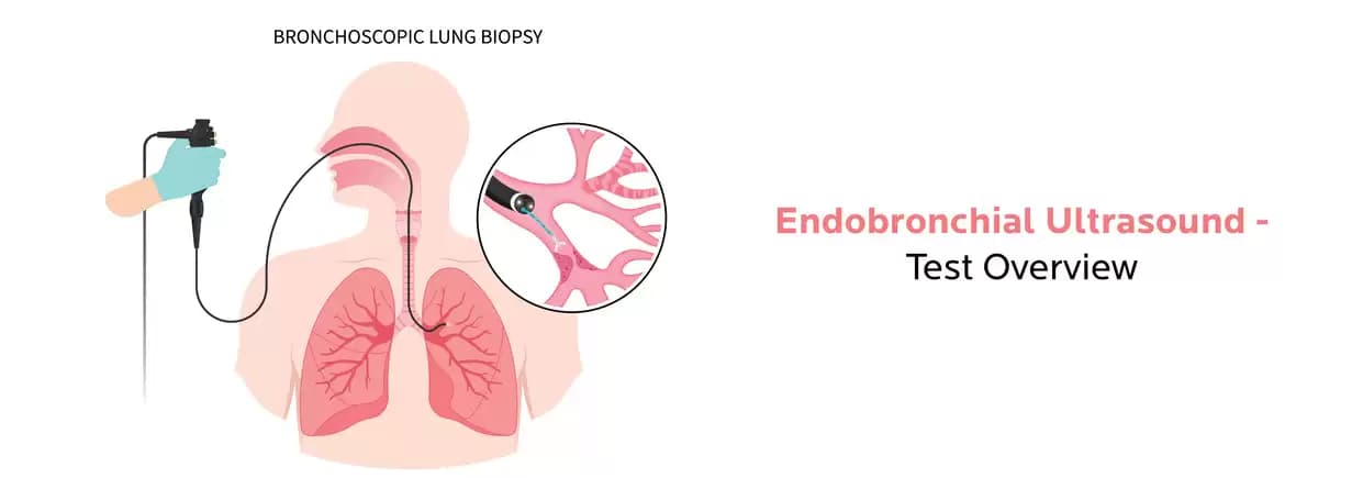
Endobronchial ultrasound (EBUS) has become an essential method for medical professionals for diagnosing, staging, and managing several lung diseases, particularly lung cancer. This minimally invasive procedure allows healthcare professionals for real-time visualisation and targeted sampling, aiding in appropriate diagnosis and planning treatment for the patient.
Endobronchial ultrasound (EBUS) has become an essential method for medical professionals for diagnosing, stage, and managing several lung diseases, particularly lung cancer. This minimally invasive procedure allows healthcare professionals for real-time visualization and targeted sampling, aiding in appropriate diagnosis and planning treatment for the patient.
EBUS averts the want for more invasive surgical procedures for tissue sampling or assessing lung and mediastinal lesions. During the EBUS procedure, the physician can involve performing fine-needle aspiration (FNA) or biopsy techniques to gather samples of suspicious tissues or lymph nodes for further evaluation.
In this blog, we will cover everything that’s related to EBUS procedure so make sure to give it a read till the end. Kindly note that it is for informational purposes only and doesn’t rule out the doctor’s consultation.
An endobronchial ultrasound (EBUS) is a medical procedure used for visualizing and evaluating structures within the airways and lungs. It combines bronchoscopy involving the insertion of a bronchoscope through the mouth or nose and into the airways using ultrasound technology.
The EBUS procedure consents physicians to evaluate the size, location, and characteristics of anomalies within the airways like tumors or nodules. It is commonly used for diagnosing lung cancer, staging lung cancer to determine the extent of its spread, and evaluating enlarged lymph nodes for the possible presence of cancer.
Endobronchial procedures offer various advantages over traditional surgeries which includes:
A special type of ultrasound during an endobronchial ultrasound (EBUS) procedure is used to assess the airways and structures within the chest. Here's what one can usually expect during an EBUS procedure:
There are certain steps followed before the EBUS test which involve:
There are various probable outcomes and subsequent steps that may happen after an Endobronchial Ultrasound (EBUS) procedure. The main course of action relies on the purpose of the procedure and the findings during the EBUS. Here is what happens after the procedure:
Endobronchial procedures have considerably advanced the respiratory medicine field by offering the options of minimally invasive procedures to diagnose, stage, and treat several airway disorders. With constant developments in technology and techniques, these procedures continue to emerge providing promising outcomes and enhanced quality of life for patients with respiratory conditions. You can book your appointment with the best pulmonologist in Jaipur at Rukmani Birla Hospital, which offers the most exceptional care to its patients.
EBUS bronchoscopy is a procedure performed for diagnosing varied types of lung disorders which include cancer, inflammation, or infections.
No, EBUS biopsy is not painful as it is a minimally advanced invasive procedure
EBUS is not risky but one of the safest procedures and there is very less risk of complications.
Written and Verified by:
.webp&w=256&q=75)
Dr. Rakesh Godara is Additional Director of Pulmonology Dept. at CK Birla Hospital, Jaipur with over 18 years of experience. He specializes in ARDS, bronchoscopic management of hemoptysis, central airway obstruction, endobronchial ultrasound, and medical thoracoscopy/pleuroscopy.
Similar Pulmonology Blogs
Book Your Appointment TODAY
© 2024 RBH Jaipur. All Rights Reserved.