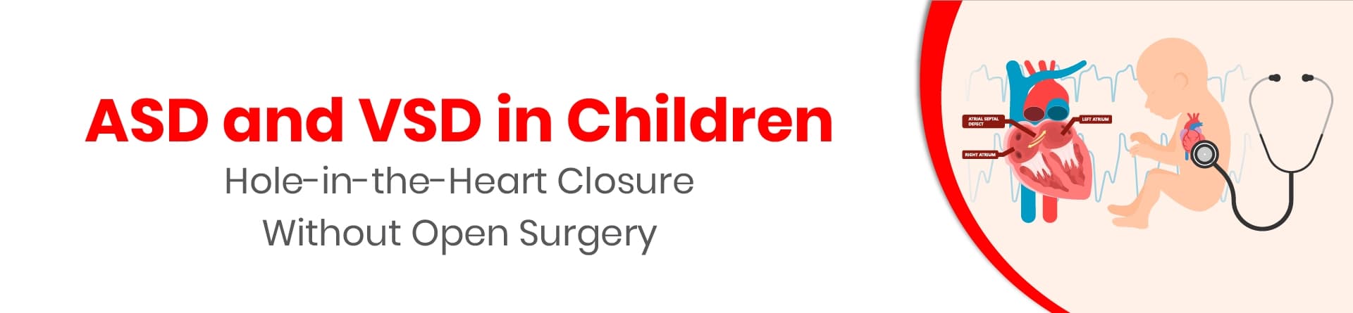
Atrial septal defect (ASD) and ventricular septal defect (VSD) are congenital heart conditions in which a child is born with an opening in the wall separating the heart chambers.
Small defects may close naturally and require regular monitoring, while larger openings may need treatment to prevent strain on the heart and lungs.
Common symptoms include breathing difficulties, poor feeding, excessive sweating during feeds, slow weight gain and frequent respiratory infections.
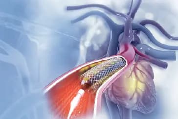
Selected ASD and VSD cases can be treated through minimally invasive device closure using a catheter, without opening the chest.
The choice between monitoring, device closure and open-heart surgery depends on the defect's size, location, effect on blood flow and the child's overall health.
Recovery after device closure is generally shorter than after open-heart surgery, although regular follow-up remains important.
A hole in the heart can sound worrying when your child is diagnosed with it. However, many children with an atrial septal defect (ASD) or ventricular septal defect (VSD) can be treated successfully. In selected cases, the hole can be closed through a minimally invasive procedure using a small device, without opening the chest.
The right treatment depends on the type, size and location of the defect, how it affects blood flow, and your child’s overall health. Some small defects may close on their own and only need regular monitoring. Others may require closure to protect the heart and lungs from extra strain.
At CK Birla Hospitals, Kolkata, our paediatric cardiac care team can assess your child’s condition and explain whether monitoring, device closure or surgery is the most suitable option.
ASD and VSD are congenital heart defects. This means a child is born with an opening in the wall, called the septum, that separates the heart’s chambers.
The heart has two upper chambers (atria) and two lower chambers (ventricles). The septum helps keep blood flowing through the heart in the right direction.
These openings can allow blood to pass between the chambers. Depending on the size and location of the defect, extra blood may flow towards the lungs, making the heart and lung blood vessels work harder.
ASD and VSD develop while the baby’s heart is forming during pregnancy. In most cases, there is no single identifiable reason for the defect.
The causes of ASD and VSD may involve a combination of genetic and environmental factors. Sometimes, these defects occur alongside other congenital heart conditions. Having a hole in the heart does not mean that a parent did anything wrong during pregnancy.
ASD and VSD in newborn babies may be detected during a routine examination, when a doctor hears a heart murmur, or when symptoms lead to further testing. An echocardiogram (heart ultrasound) is commonly used to confirm the diagnosis and assess the defect.
The symptoms of ASD and VSD vary. Some children have no noticeable symptoms, especially when the opening is small. Larger defects may cause signs that become more apparent as the baby grows.
Possible symptoms include:
A child may have one or several symptoms, or none at all. If your child has difficulty breathing, struggles to feed, or is not gaining weight as expected, seek medical assessment.
Not every ASD or VSD needs immediate treatment. The paediatric cardiologist considers the defect’s size and position, the amount of blood passing through it, and whether it is affecting the heart or lungs.
Monitoring may be suitable when the defect is small, does not place significant strain on the heart, and may close naturally. Your child may need regular check-ups and echocardiograms to monitor changes.
Closure may be recommended when the opening allows too much blood to flow to the lungs or causes enlargement or strain of the heart chambers. Treatment may also be considered when a child has symptoms such as breathing difficulty, poor feeding or slow weight gain.
The timing of treatment is individual. A large defect may need early intervention, while a smaller one may be monitored for longer. The goal is to prevent complications while choosing the safest approach for your child.
Device closure is a minimally invasive procedure performed through cardiac catheterisation. Instead of making an incision in the chest, the doctor guides a thin, flexible tube called a catheter through a blood vessel, usually in the groin, towards the heart.
A small closure device is passed through the catheter and positioned across the opening. Once the doctor confirms that it is correctly placed, the device is released to close the defect. Over time, the child’s heart tissue grows over the device.
The procedure is generally performed under anaesthesia, with the team using imaging such as X-ray and echocardiography to guide placement.
Device closure is commonly used for a suitable secundum ASD, which is a defect in the central part of the wall between the upper chambers.
The cardiologist checks whether the opening has the right size and enough surrounding tissue to hold the device securely. Other ASD types may require surgical repair instead.
Some VSDs can be closed using a catheter-based device, but this approach is suitable only for selected defects. The location of the opening and its relationship to nearby heart valves and other structures are important.
Open-heart surgery remains the standard approach for many VSDs. Your child’s cardiologist will explain whether device closure is an appropriate option or whether surgery offers a safer and more effective repair.
Device closure is not suitable for every child or every type of ASD and VSD. The treatment plan is based on detailed assessment rather than the diagnosis alone.
Your child may be considered for device closure if:
Before recommending treatment, the paediatric cardiology team may perform an echocardiogram, ECG and other tests as needed. The team will also assess your child’s age, weight, symptoms and overall health.
For some children, open-heart surgery may be the more appropriate option. The cardiologist will discuss the benefits and risks of each approach with you.
Recovery after device closure is usually shorter than recovery after open-heart surgery because the chest is not opened. However, the length of hospital stay and return to usual activities depend on your child’s condition and the hospital’s care plan.
After the procedure, your child will be monitored for heart rhythm, breathing, blood pressure and the catheter insertion site. The team may perform an ECG or echocardiogram to check the device’s position and assess blood flow.
Many children go home the same day or the following day if they are stable. At home, you may be advised to:
Your child’s doctor will tell you when they can return to school, sports and other activities. Follow-up care remains important even when your child feels well.
Written and Verified by:

Dr. Satarupa Mukherjee is an Consultant in Pediatric Cardiology at BM Birla Heart Hospital, Kolkata with over 13 years of experience. She specializes in post-op care after neonatal & pediatric cardiac surgery, extracorporeal life support, and pediatric cardiac transplantation.
Similar Pediatric Cardiology Blogs
Book Your Appointment TODAY
© 2024 BMB Kolkata. All Rights Reserved.